A PSMA PET scan is a specialised PET-CT imaging test primarily used for the detection, staging, and monitoring of prostate cancer.
Unlike routine PET scans, PSMA PET imaging targets Prostate-Specific Membrane Antigen (PSMA), which is highly expressed on prostate cancer cells.
This advanced scan is performed only when clinically indicated and is recommended by doctors for accurate disease assessment and treatment planning.
Patients from Chandigarh, Mohali, and Panchkula (Tricity) are referred for PSMA PET scans when precise and targeted imaging is required for prostate cancer evaluation.
Understanding PSMA-Targeted PET Imaging
A PSMA PET scan is a receptor-targeted nuclear medicine imaging test designed to identify prostate cancer cells with high sensitivity. It works by using a radiotracer that binds specifically to PSMA receptors present on prostate cancer tissue.
Because of this targeted mechanism, PSMA PET scans can detect disease even at very low PSA levels, making them significantly more accurate than conventional imaging in many clinical scenarios.
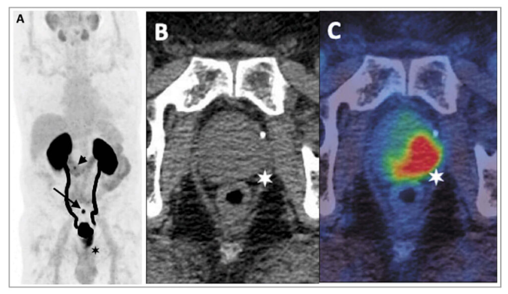
Role of Ga-68 PSMA PET in Prostate Cancer ImagingGa-68 PSMA
PET imaging uses a Gallium-68 labelled radiotracer that binds selectively to PSMA receptors. After administration, the tracer circulates through the body and attaches to prostate cancer cells, thereby enabling high-resolution PET-CT visualisation.
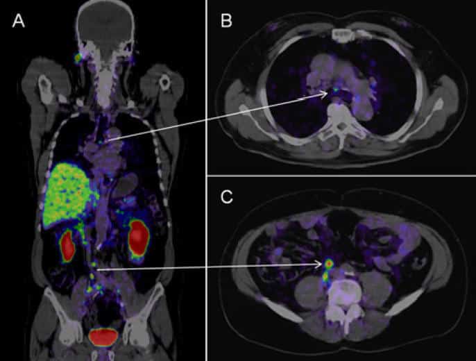
As a result, Ga-68 PSMA PET scans are widely used for:
- Detecting recurrent prostate cancer
- Identifying metastatic spread
- Supporting treatment planning and monitoring
How PSMA PET Scan Differs from Conventional PET Imaging
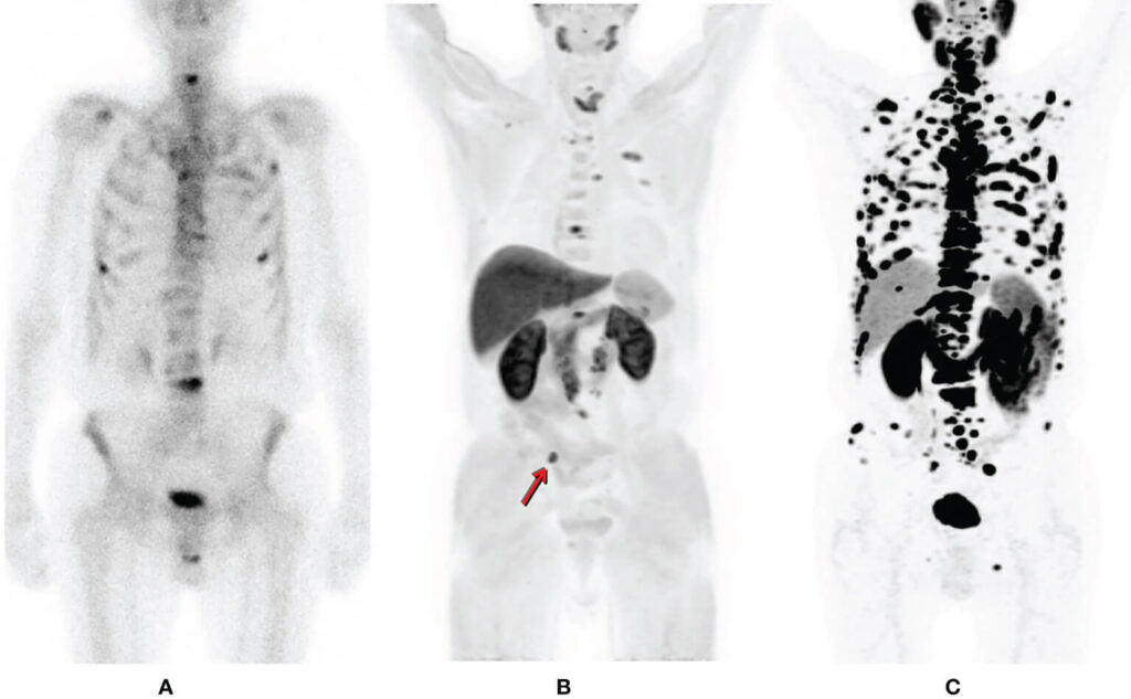
Although both are PET-CT scans, PSMA PET and FDG PET serve different purposes. FDG PET imaging measures glucose metabolism and is commonly used for many cancers. However, prostate cancer often shows limited FDG uptake.
In contrast, PSMA PET imaging focuses specifically on prostate cancer cells. Therefore, it provides greater sensitivity and specificity for prostate cancer evaluation, particularly in early recurrence and low PSA scenarios.
Clinical Situations Where PSMA PET Scan Is Recommended
Doctors may recommend a PSMA PET scan in several situations, including:
- Initial staging of prostate cancer
- Evaluation of biochemical recurrence
- Detection of metastatic disease
Assessment before targeted or systemic therapy
Because of its targeted nature, PSMA PET imaging helps clinicians make informed decisions with confidence.
Step-by-Step: How a PSMA PET Scan Works
First, a PSMA-targeted radiotracer is administered intravenously. Next, the tracer binds to prostate cancer cells expressing PSMA receptors. Subsequently, PET-CT imaging captures detailed functional and anatomical images.
Together, these images allow doctors to accurately identify disease location, extent, and progression, which is essential for effective treatment planning.
Preparation Guidelines for PSMA PET Scan
Preparation for a PSMA PET scan is usually simple. However, patients are advised to carefully follow instructions provided by the referring doctor or imaging centre. This may include guidance related to fasting, medications, or prior investigations.
By following preparation guidelines, patients help ensure reliable imaging and accurate interpretation.
Diagnostic Accuracy and Clinical Value of PSMA PET Imaging
PSMA PET imaging is recognised for its high diagnostic accuracy in prostate cancer evaluation. Because it directly targets cancer-specific receptors, it reduces uncertainty and improves lesion detection.
Furthermore, PSMA PET results play a key role in guiding treatment decisions, evaluating therapy response, and planning follow-up care, thereby improving overall clinical outcomes.
Why Medical Referral Is Essential for PSMA PET Scan
A PSMA PET scan is performed only when clinically justified. Therefore, a doctor’s referral is essential to confirm the indication, select the appropriate imaging protocol, and ensure safe tracer use.

Additionally, medical guidance is required to correctly interpret scan findings and integrate them into diagnosis, treatment planning, and long-term disease monitoring.
PSMA PET Scan Services Across Tricity
Patients from Chandigarh, Mohali, and Panchkula commonly undergo PSMA PET imaging when specialised prostate cancer evaluation is required. This scan is typically recommended by urologists, oncologists, or nuclear medicine specialists as part of comprehensive cancer care.

For routine PET-CT imaging and general diagnostic evaluation, patients may refer to our detailed PET Scan in Chandigarh information page.
Frequently Asked Questions About PSMA PET Scan
What is a PSMA PET scan?
A PSMA PET scan is a specialised PET-CT imaging test used to detect and evaluate prostate cancer. It works by targeting Prostate-Specific Membrane Antigen, a protein commonly found on prostate cancer cells, allowing precise identification of disease location and spread.
What is a PSMA PET scan used for?
PSMA PET scans are used for prostate cancer staging, detecting recurrence, identifying metastasis, and guiding treatment planning. Doctors recommend this scan when detailed and targeted imaging is required for accurate clinical decision-making.
How is PSMA PET scan different from FDG PET scan?
A PSMA PET scan targets prostate cancer cells specifically, while an FDG PET scan measures general glucose metabolism. Because prostate cancer often shows low FDG uptake, PSMA PET imaging provides higher accuracy for prostate cancer evaluation.
What is Ga-68 PSMA PET imaging?
Ga-68 PSMA PET imaging uses a Gallium-68 labelled radiotracer that binds to PSMA receptors on prostate cancer cells. This allows doctors to detect cancer even at low PSA levels and assess disease extent with high precision.
How accurate is a PSMA PET scan?
PSMA PET scans are highly accurate for detecting prostate cancer due to their receptor-specific targeting. They are especially effective in identifying early recurrence and small metastatic lesions that may not be visible on conventional imaging.
Does a PSMA PET scan require a doctor’s prescription?
Yes, a PSMA PET scan requires a referral from a licensed doctor. Since it is a specialised diagnostic test, medical recommendation ensures the scan is appropriate and that results are correctly interpreted for treatment planning.
Is special preparation needed before a PSMA PET scan?
Preparation for a PSMA PET scan is usually minimal. Patients are typically advised to inform the medical team about medications and previous treatments and follow the specific instructions provided by the referring doctor.
