Best Diagnostic Center in Chandigarh for All Medical Imaging Needs
As the leading diagnostic imaging center in Chandigarh, we provide comprehensive radiology services including MRI, CT Scan, PET-CT, Ultrasound, X-ray, Echocardiography, ECG, and EEG. Our state-of-the-art facilities are equipped with the latest diagnostic technology to deliver accurate results for patients across Chandigarh, Mohali, and Panchkula.
Why Choose Our Diagnostic Services in Chandigarh?
- Advanced technology with 1.5T and 3.0T MRI scanners
- Most affordable rates for all diagnostic tests in Chandigarh Tricity
- Same-day reporting by specialized radiologists
- Conveniently located centers across Chandigarh
- Complimentary pick and drop services for patients
- Open 7 days a week including holidays
Visit our centers in Sector 34, Sector 22, or Sector 17 Chandigarh for the best diagnostic experience. Book your appointment today for high-quality, affordable diagnostic services in Chandigarh.
Here’s a bucket of medical information on: How can brain tumors be diagnosed?
Your brain is the most vital organ in your body that controls & regulates the functions of all the others!
And,
A brain tumor, be it benign or malignant, is a serious threat to topple!
So,
Multidisciplinary care stands requisite herein! With the advancement of medical science & technologies, diagnosing brain tumors has turned efficient & easy!
And,
Your doctor shall accommodate all tests & approaches possible altogether to offer you an early diagnosis!
Typically, the diagnosis of a brain tumor starts with three approaches; the physical exam, the neurological exam, and the visual field exam!
Physical Exam
This approach includes a study of your body from the outside to check the signs and symptoms you bear. Your doctors shall touch the head skin and try to feel it if they can locate a lump-like structure therein!
During this exam, your doctors shall ask you various questions regarding your health history, your family health history, the medications you intake, your age, workspace, lifestyle habits, and much more!
Neurological Exam
The next approach to follow is neurological testing! This is where a series of tests shall take place to see the condition of your nerves, mental status, coordination between your brain, spinal cord, & muscles, how well your senses and reflexes work, and so forth!
During this exam, your doctors may shoot a list of questions related to your movements, your memories, and your behaviors! Your doctors want to check whether your cognitive processes are on track alongside your neurological functions!
Visual Field Exam
Then comes another diagnostic approach called the Visual Field Exam! This test is where your vision field, i.e., the total area in which you can see various objects, shall lay assessed! If they find a loss of vision, they consider it a sign of a tumor that caused damage to the part of the brain affecting eyesight.
In this regard, your doctors are likely to assess both your central vision & peripheral vision. Thus, they can understand how much you can see when looking straight and how much you can see in all other directions again when you look straight!
Now,
After this round,
Following the test results, when your doctor predicts a tumor growing beneath your skin & flesh, you shall have to go through a series of body scans or imaging tests. It is because your doctors need to confirm the case and get an in-depth understanding of – How big the tumor is, where it exactly resides, what shape it has, and whether there is one tumor or many!
The imaging tests that your doctor may order are as follows!
MRI with Contrast
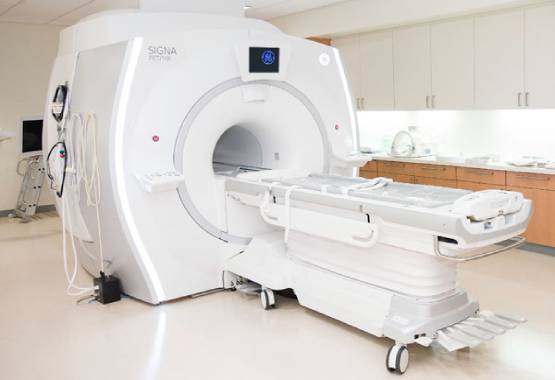
This is the diagnostic process in which strong magnets lay used alongside radio waves & a computer console to create precise internal imaging of your brain. During this test, you shall receive the gadolinium-based contrast agents intravenously to ensure better clarity of the imaging and locate the brain tumor, even if it is tiny in size.
It stands for Magnetic Resonance Imaging Test, the one safest in radiology because of non-invasion and non-radiation!
Advanced MRI
This is another of the Magnetic Resonance Imaging approach conducted to see the tumor’s proximity to a critical part of your brain. This test, being an advanced technique of MRI, can trace & determine multiple other characteristics of the brain tumor so that the medical caregivers can plan the best possible treatment.
Advanced MRI approaches are of different types, namely –
- Functional MRI, where your blood flow gets assessed along with the parts of your brain responsible for performing critical functions,
- Diffusion Tensor Imaging or DTI, where the white matter in your brain gets assessed to trace the cellularity, structure, and nature of the brain tumor; used mainly during preoperative planning,
- Perfusion MRI, where the blood volume & grade of your brain tumor lay assessed,
- Hemosiderin Imaging, where the presence of occult blood in the brain if any, lay traced,
- Magnetic Resonance Spectroscopy or MRS, where the biochemical changes in your brain during the presence of a tumor lay measured, particularly to see the progression of the tumor and its metabolism! This approach serves to monitor & regulate a tumor removal treatment!
CT Scan
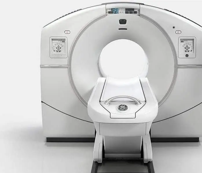
CT scan, known as Computed Tomography Scan in complete terms, is the diagnostic approach in which an X-Ray machine and a computer lay used to create a series of imaging of your brain. It may also be referred to as computerized tomography or computerized axial tomography!
During this test, you are injected with ionizing radiation through an intravenous needle into your veins, which makes the tumor appear bright on the computer screen. Sometimes, the radioactive materials may be administered orally like a pill to swallow!
PET Scan
PET scan, or Positron Emission Tomography Scan, is the diagnostic process in which a rotating machine moves around your body while radioactive glucose injected into your veins traces the growing brain tumor!
This scan is more used to detect a malignant tumor, as the radioactive glucose lay absorbed by the cancerous cells more than a benign mass. It helps the doctor understand whether it is a tumor in real or just an Inflammation!
Diagnostic Angiogram
This is the diagnostic approach in which your blood vessels & blood flow lay assessed mainly, and your doctor orders this test to see if any of the blood vessels lay blocked because of the growing tumor! The machine used here is the same X-Ray machine used for a usual body scan!
This process may or may not involve the use of contrast dye!
Bottom Line:
Above all,
If the imaging tests confirm that you have a brain tumor, a biopsy shall be conducted thereupon!
For availing of an MRI to diagnose a brain tumor, you can feel free to visit www.mrichandigarh.com!


