
Description
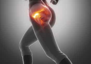
A Contrast CT Scan of the Hips is an advanced imaging test used to examine the hip joints, pelvic bones, muscles, blood vessels, and surrounding soft tissues. Because a contrast dye is injected before the scan, abnormal areas become more visible, and therefore doctors can detect disease with greater precision.
Moreover, by combining contrast enhancement with high-resolution CT imaging, this test provides a much clearer picture of hip-related problems.
What Is a Contrast CT Hips Scan?
A contrast-enhanced CT of the hips uses X-ray technology and computer processing to generate detailed cross-sectional images. As a result, both bone and soft-tissue structures can be evaluated accurately, which consequently improves diagnosis.
Why Is a Contrast CT of the Hips Performed?
This scan is recommended when symptoms are persistent or severe, and therefore it is used to:
- Therefore, detect hip joint infections or inflammation
- Additionally, identify tumors or abnormal growths
- Moreover, evaluate blood vessel problems
- Furthermore, assess injuries or fractures
- Finally, plan surgery or monitor treatment response
How Is the Scan Performed?
Before the scan, a contrast dye is injected into a vein. Meanwhile, you will lie on the CT scanner table while images of the hips are taken. Consequently, the scanner produces detailed pictures within minutes.
Most importantly, the test is painless and usually takes only 10 to 15 minutes.
Benefits of Contrast CT Hips Imaging
- Firstly, it provides detailed views of hips and pelvis
- Secondly, it detects hidden disease
- Thirdly, it improves diagnostic confidence
- In addition, it supports accurate treatment planning
- Overall, it leads to better patient care
Book a Contrast CT Hips Scan
If you have persistent hip pain, swelling, or mobility issues, a Contrast CT Hips Scan not only clarifies the cause but also helps your doctor choose the most effective treatment.


Comments