Best Diagnostic Services in Chandigarh
Premier Radio Diagnostic Center in Chandigarh TricityWe are the leading diagnostic centre in Chandigarh providing comprehensive radio diagnostic services including MRI, CT Scan, PET Scan, Ultrasound, X-ray, Echocardiogram, ECG, and EEG at affordable rates with same-day reporting.
Best Diagnostic Center in Chandigarh for All Medical Imaging Needs
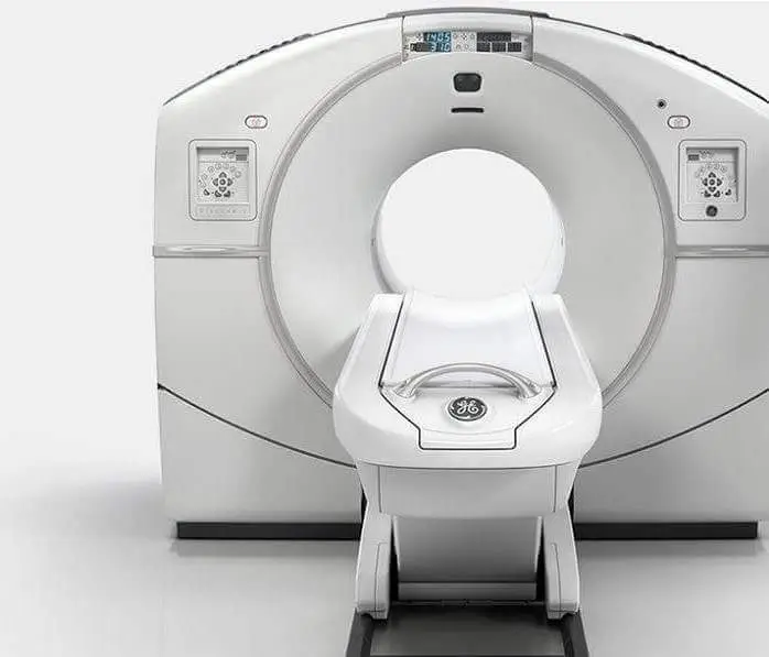
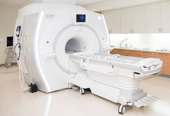
As the leading diagnostic imaging center in Chandigarh, we provide comprehensive radiology services including MRI, CT Scan, PET-CT, Ultrasound, X-ray, Echocardiography, ECG, and EEG. Our state-of-the-art facilities are equipped with the latest diagnostic technology to deliver accurate results for patients across Chandigarh, Mohali, and Panchkula.
Why Choose Our Diagnostic Services in Chandigarh?
- Advanced technology with 1.5T and 3.0T MRI scanners
- Most affordable rates for all diagnostic tests in Chandigarh Tricity
- Same-day reporting by specialized radiologists
- Conveniently located centers across Chandigarh
- Complimentary pick and drop services for patients
- Open 7 days a week including holidays
Visit our centers in Sector 34, Sector 22, or Sector 17 Chandigarh for the best diagnostic experience. Book your appointment today for high-quality, affordable diagnostic services in Chandigarh.
Can MRI scan detect heart problems?
MRI can be used to image the heart and blood vessels and can be used to detect a variety of heart problems. Some of the ways that are used to evaluate heart function and structure include:
- Identifying problems with the heart’s valves, such as stenosis (narrowing) or regurgitation (leakage).
- Evaluating the heart muscle and blood vessels to detect problems such as heart attacks, inflammation of the heart muscle (myocarditis), or blockages in the coronary arteries (the vessels that supply blood to the heart muscle).
- Detecting and assessing the size and location of any tumors or other abnormal growths in the heart or blood vessels.
- Identifying problems with the heart’s electrical conduction system, which can cause arrhythmias (abnormal heart rhythms).
- Assessing blood flow in the heart and blood vessels and identifying any areas of restricted or increased flow.
- It’s important to note that MRI is only a diagnostic tool, and the interpretation of the images obtained and the diagnosis of heart disease is done by a Cardiologist. and further tests may be needed to confirm the diagnosis or treatment options.
MRI scans are a powerful tool for detecting various medical conditions and illnesses, including heart problems. An MRI scan is a painless test that uses powerful magnets and radio waves to create detailed images of the inside of your body. It can help diagnose cardiovascular issues such as blocked arteries or an enlarged heart without the need for invasive surgery.
The MRI scan works by creating a high-resolution image of your heart, allowing doctors to look for signs of damage or blood clots. It can also detect other abnormalities, such as tumors or cysts in the heart muscle and chambers. In addition, it can provide insight into how well your heart valves are functioning and whether they need repair or replacement.
Overall, an MRI scan is an invaluable tool when it comes to detecting potential heart problems before they become serious issues.
You can call us for More Information, at 8699572364
Follow us on Social Pages: mrichandigarh
Instagram: Mri Chandigarh
Mail us at [email protected]
Tweet: Mri Chandigarh
Linked In: Mri Chandigarh


