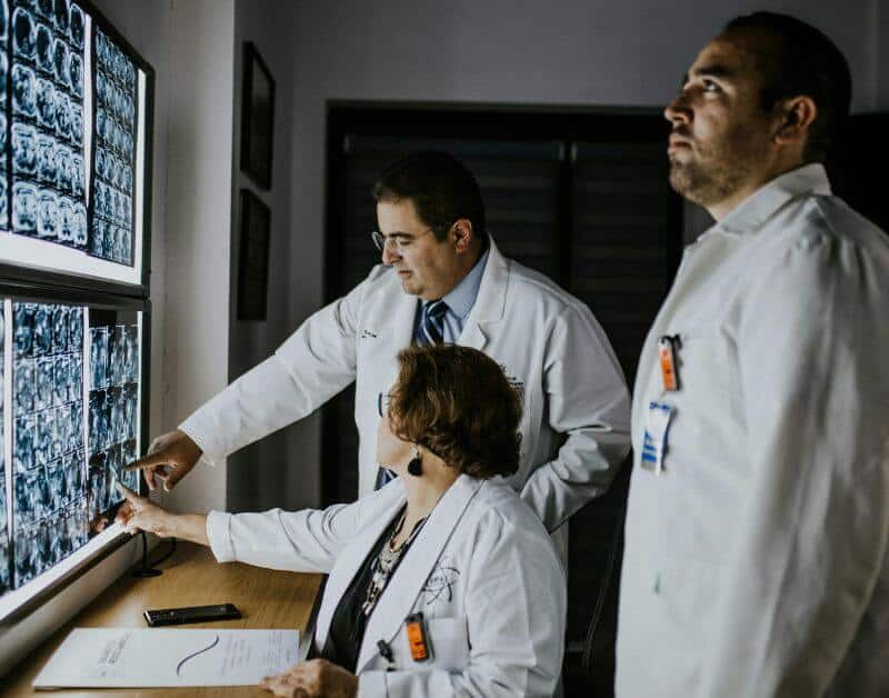
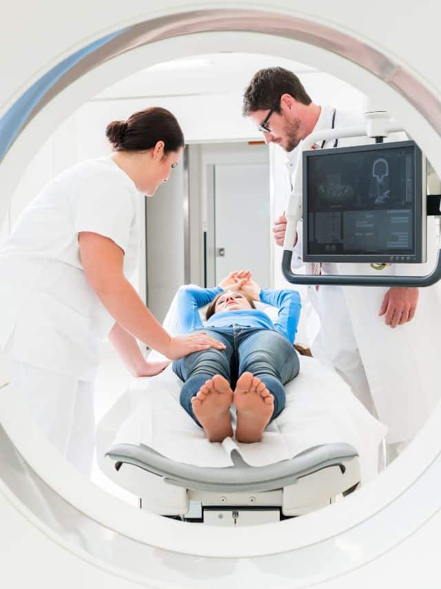
We have in-house Radiologist at MRI Chandigarh so, you will get same day report with accurate results.
We provide free pick and drop facility for our patients in tricity for MRI, CT Scan, PSMA PET Scan, DOTA PET Scan and PET Scan @ ₹12,999 in Chandigarh.
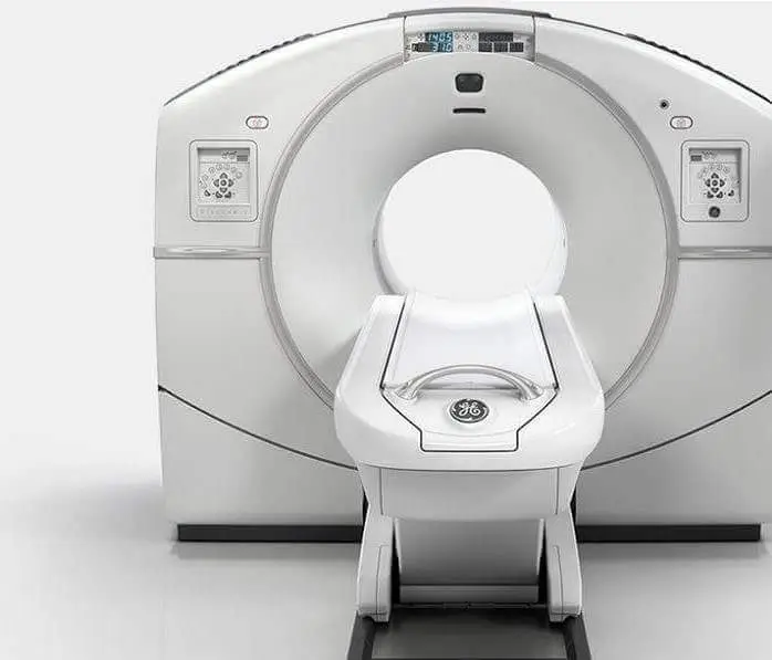
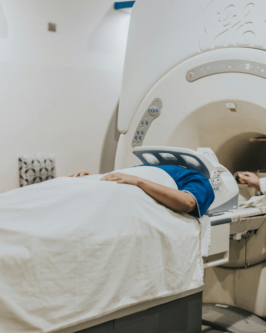
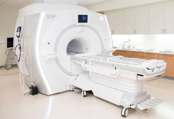
We are equipped with World Class MRI Scan Facility at Reasonable pricing. With best in class skilled technicians.
Get a comfortable MRI scan in Chandigarh using high-quality 1.5T and 3T machines with expert reporting and transparent, patient-friendly pricing.
MRI Chandigarh is your one-stop destination for advanced medical imaging and diagnostic services in Chandigarh. As one of the most trusted MRI centres in Chandigarh, we provide cutting-edge radio-imaging solutions, including both 1.5 Tesla and 3 Tesla MRI scans, at highly competitive prices—without compromising on quality.
We offer quick, accurate, and affordable MRI scans in Chandigarh with no hidden charges. Our experienced radiologists ensure timely results and, if needed, offer recommendations for further medical consultation based on your MRI report.
The cost of an MRI scan in Chandigarh varies depending on the type of scan and the diagnostic center. Prices typically start from ₹3,800 to ₹5,500. For example, 1.5 Tesla MRI scans may begin at ₹3,800, while 3 Tesla MRI scans are priced higher due to their advanced imaging capabilities. It's advisable to contact local MRI centers for precise pricing.
Preparation for an MRI scan generally involves minimal steps. For most MRI scans, you can maintain your regular diet. However, for abdominal scans, fasting for 3-4 hours prior to the procedure may be required. Wear loose-fitting, metal-free clothing, and inform your radiologist about any existing medical conditions, allergies, or implants.
Yes, MRI scans are generally considered safe as they do not use ionizing radiation. However, patients with certain implants like pacemakers should inform their doctor beforehand. It's crucial to remove all metal objects before the scan to prevent any risks associated with the MRI's strong magnetic field.
The duration of an MRI scan depends on the area being examined but typically ranges from 15 minutes to over an hour. For instance, a brain MRI may take about 30 to 60 minutes, while joint MRIs like knee or shoulder can take 15 to 45 minutes. Advanced 3 Tesla MRI machines can perform scans faster, enhancing patient comfort.
Chandigarh hosts several reputable MRI centres equipped with state-of-the-art technology and experienced radiologists. Centers like MRI Chandigarh are known for their advanced imaging services, including 1.5 Tesla and 3 Tesla MRI scans, and patient-centric care. It's recommended to research and choose a center that best fits your diagnostic needs.
While some diagnostic centers may allow self-referred MRI scans, it's advisable to consult with a healthcare professional to ensure the appropriateness of the scan and to provide the necessary clinical context for accurate interpretation.
Yes, certain MRI centres in Chandigarh offer complimentary pick-up and drop-off services within the Tricity area (Chandigarh, Mohali, Panchkula) to enhance patient convenience. It's best to inquire with the specific MRI center about the availability of such services. We provide free pick and drop in tricity for all scans including CT Scan & PET Scan too.
We are the leading diagnostic centre in Chandigarh providing comprehensive radio diagnostic services including MRI, CT Scan, PET Scan, Ultrasound, X-ray, Echocardiogram, ECG, and EEG at affordable rates with same-day reporting.
Experience the most comfortable and advanced MRI scans in Chandigarh with our high-field strength 1.5 Tesla and 3.0 Tesla machines. Our state-of-the-art MRI technology delivers exceptional quality diagnostic images for accurate results. Located in Sector 34, we provide the most reliable MRI services in Chandigarh Tricity at affordable rates.
Our Chandigarh diagnostic center provides world-class CT Scan facilities at the most competitive prices in the tricity region. Equipped with low-radiation dose CT scanners and operated by skilled technicians, we ensure the safest and most accurate CT scan services in Chandigarh, Mohali and Panchkula area.
Get the most affordable PET scan in Chandigarh at just Rs.16,000, the lowest price in the tricity region. Our all-inclusive package offers free pickup and drop, complimentary meals, and patient comfort amenities. Our Chandigarh PET scan center uses state-of-the-art technology for early cancer detection and treatment monitoring.
Our Chandigarh diagnostic center offers high-precision ultrasound services including 2D, 3D, and 4D scans. We use latest technology equipment at our partner diagnostic facilities across Chandigarh Sectors 34, 35, and 22. Our experienced radiologists provide accurate results for abdominal, pregnancy, thyroid, and other specialized ultrasound examinations.
Our network of partner X-ray centers in Chandigarh offers affordable digital X-ray services with instant results. Using advanced digital radiography technology, we provide high-quality bone and soft tissue imaging with minimal radiation exposure. Visit our X-ray diagnostic facilities in Sectors 34, 22, and 17 of Chandigarh for quick and reliable service.
Our diagnostic center in Chandigarh provides comprehensive cardiac and neurological testing including Echocardiography (Echo), Electrocardiogram (ECG), and Electroencephalogram (EEG). We offer the most affordable rates for these specialized tests in the Chandigarh region with reports prepared by experienced cardiologists and neurologists for accurate diagnosis.
As the leading diagnostic imaging center in Chandigarh, we provide comprehensive radiology services including MRI, CT Scan, PET-CT, Ultrasound, X-ray, Echocardiography, ECG, and EEG. Our state-of-the-art facilities are equipped with the latest diagnostic technology to deliver accurate results for patients across Chandigarh, Mohali, and Panchkula.
Visit our centers in Sector 34, Sector 22, or Sector 17 Chandigarh for the best diagnostic experience. Book your appointment today for high-quality, affordable diagnostic services in Chandigarh.
FDG-DOPA PET scans are increasingly being used in the diagnosis and management of neurodegenerative diseases. The aim of this study was to evaluate the feasibility and accuracy of FDG-DOPA PET in the diagnosis of Parkinson’s disease (PD) and dementia with Lewy bodies (DLB) in a tertiary care center in North India. A total of 15 patients with a clinical diagnosis of PD, 10 patients with a clinical diagnosis of DLB, and 15 healthy controls were included in this study. All participants underwent FDG-DOPA PET imaging. The images were analyzed for standardized uptake value (SUV) ratios and visual interpretation by two nuclear medicine physicians blinded to the clinical diagnosis. The SUV ratios were significantly higher in patients with PD and DLB as compared to healthy controls (P < 0.001).
FDG DOPA PET scans are used to diagnose and monitor tumors. The combination of FDG and DOPA is especially useful for identifying small lesions in the brain. A recent study found that the use of FDG DOPA PET scans in Chandigarh is reliable for diagnosing and monitoring tumors.
FDG DOPA PET scans are a valuable tool for diagnosing and staging various cancers. The use of FDG DOPA PET scans is increasing in many parts of the world, including Chandigarh. FDG DOPA PET scans are very helpful in determining the extent of cancer and whether it has spread to other parts of the body. They can also help doctors determine the best treatment plan for a patient.
FDOPA PET Scan is the best test to detect early Parkinson’s disease. It is a simple, painless, and non-invasive test. The scan uses a small amount of radioactive material called F-18 fluorodeoxyglucose (FDG) which is injected into a vein and taken up by the brain cells.
The Chandigarh region has one of the highest incidences of Parkinson’s disease in the world. The Department of Neurology at Government Medical College and Hospital (GMCH), Chandigarh, has started offering FDG DOPA PET Scan to diagnose early Parkinson’s disease.
In Chandigarh, FDG DOPA PET scans are used to diagnose cancer and other diseases. The scan is used to measure the level of dopamine in the brain. This helps doctors determine if a patient has a tumor or other disease that is affecting the dopamine levels in the brain.
An FDG DOPA PET scan is a nuclear medicine test that uses a small amount of radioactive material, called a tracer, to look at how well the brain is working. The tracer is injected into a vein and travels through the body to the brain. There it binds to certain cells in the brain that use dopamine. A special camera is used to take pictures of the tracer in the brain. These pictures can show how well the cells are working and help doctors decide if treatment is working.
FDG DOPA PET scans are used to help doctors determine how well treatments for Parkinson’s disease are working. This type of scan can also help doctors see if a person’s symptoms are getting worse. The scans use a small amount of radioactive material that is injected into a vein. This material helps create images of the brain.
There are many potential reasons why a doctor might ask for an FDG DOPA PET scan. One reason could be to help diagnose Parkinson’s disease. This is a neurological disorder that affects movement and can cause tremors and other problems. An FDG DOPA PET scan can help doctors see how much of the brain is affected by the disease and can help them plan treatment.
Another reason a doctor might order an FDG DOPA PET scan is to check for tumors in the brain. Tumors can affect how the brain functions, so it’s important to find them as early as possible. An FDG DOPA PET scan can help doctors see if there are any tumors in the brain and how big they are.
An FDG DOPA PET Scan is a nuclear medicine procedure that uses a radioactive tracer called fluorodeoxyglucose (FDG) to look at how well your brain cells are working. The scan is used to help diagnose and treat conditions such as:
-Dementia
-Parkinson’s disease
-Stroke
-Brain tumor
-Epilepsy
An FDG DOPA PET Scan is a nuclear medicine study that uses a radioactive tracer called fluorodeoxyglucose (FDG). The tracer is injected into a vein and travels through the body. It is taken up by cells that use sugar (glucose) for energy, such as cancer cells. A PET scanner images the radiation is given off by the tracer. This lets your doctor see how well the tumor is responding to treatment.
There are some risk factors associated with FDG DOPA PET Scan that you should be aware of before consenting to this test:
1) You may have an allergic reaction to the radioactive material used in the scan.
2) The amount of radiation you are exposed to during the scan is slightly more than what you are normally exposed to in everyday life. However, this exposure is still considered safe.
An FDG DOPA PET scan is a nuclear medicine examination that uses a radioactive tracer called fluorine-18 fluorodeoxyglucose (FDG) to look at the brain. FDG is injected into a vein and travels through the body. It is taken up by cells that use sugar (glucose) for energy, such as cancer cells and nerve cells in the brain. A special camera takes pictures of where FDG has collected in the body. These pictures can show how well the brain is working and whether or not there are any signs of cancer.
FDG DOPA PET Scan is a nuclear medicine test that is used to help diagnose and treat certain medical conditions. The test uses a small amount of radioactive material, called a tracer, to help locate and measure the function of specific organs or tissues in the body. An FDG DOPA PET Scan is typically performed by injecting a radioactive tracer called fluorodeoxyglucose (FDG) into a vein. The tracer travels through the body and is taken up by cells that use glucose for energy. The PET scanner then creates images that show where the FDG was taken up in the body.
The FDG DOPA PET Scan is a nuclear medicine test that helps in the diagnosis of Parkinson’s disease. The scan uses a radioactive tracer called fluorodeoxyglucose (FDG), which is injected into a vein. The tracer attaches to dopamine-producing cells in the brain. A special camera then takes pictures of the brain, which are used to create a 3-D image. This image can help doctors see how well the medication is working and whether the patient needs more treatment.
FDG DOPA PET Scan is a nuclear medicine imaging test that uses a radioactive drug called fluorodeoxyglucose (FDG) and a special camera to create pictures of the body. The camera detects the radiation given off by FDG as it moves through tissues in the body. FDG is taken up by cells that use sugar (glucose) for energy, such as cancer cells.
For this test, you will be injected with FDG. You will then wait an hour for the drug to circulate through your body. You will then be placed in the scanner and pictures will be taken. The scan usually takes about 30 minutes.
A DOPA PET scan is a type of brain scan that uses a special dye called FDOPA to help doctors see how well the brain is working. This type of scan is often used to diagnose problems with the brain such as Parkinson’s disease. The dye is injected into a vein and travels through the body to the brain. A scanner then takes pictures of the brain.
A PET scan is a nuclear medicine test that uses a small amount of radioactive material, called a tracer, to help evaluate organ function and structure. The tracer is injected into a vein and travels through the body. PET scans can be used to detect problems in many organs, including the brain.
FDOPA PET scans are used to help diagnose and track the progression of Parkinson’s disease. In people with Parkinson’s, the FDOPA PET scan will show decreased uptake of the tracer in certain areas of the brain. This decrease in uptake is associated with the loss of dopamine-producing cells in the brain which is characteristic of Parkinson’s disease.
A PET scan can help to diagnose Parkinson’s disease by looking at the level of dopamine in the brain. Dopamine is a chemical that helps to control movement. A PET scan can also help to monitor the progression of the disease. The scan uses a radioactive tracer which is injected into the patient. The tracer attaches to dopamine receptors in the brain. The scanner then creates images of the brain which can show where there is a high or low concentration of dopamine receptors. This can help to determine how severe the disease is and how it is progressing.
FDG uptake on a PET scan is a measure of how much of the radioactive tracer FDG has been taken up by the body’s tissues. The more FDG that is taken up, the brighter the image on the PET scan will be. This can help doctors to determine whether or not there is a tumor present in the body.
A PET scan is a nuclear medicine test that uses a small amount of radioactive material, called a tracer, to look at how organs and tissues are working. The radioactive material is injected into a vein in your arm and travels through your body.
The tracer attaches to cells in the organ or tissue that is being studied. A scanner detects radioactivity and creates pictures on a computer screen.
PET scans can be used to detect problems with the brain, such as damage from a stroke or tumor. They can also be used to measure the amount of glucose (sugar) in the brain. This can help doctors diagnose and monitor diabetes, Alzheimer’s disease, and other conditions.
Recently, PET scans have been used to measure levels of dopamine in the brain. Dopamine is a chemical that helps control movement.
PET scans are used to detect a variety of brain disorders. Conditions that can be diagnosed with a PET scan include Alzheimer’s disease, Parkinson’s disease, and various types of cancer. The most common use for PET scans is in the detection of cancer, but they can also be used to diagnose other medical conditions.
A PET scan uses a small amount of radioactive material that is injected into the patient’s bloodstream. This material is absorbed by the cells in the body, including the cells in the brain. A scanner then measures the amount of radiation emitted by the cells. This information is used to create images of the brain that can be used to diagnose medical conditions. A PET scan is a type of imaging test that uses radiation to create pictures of the inside of the body. A PET scan can be used to detect a number of different brain disorders, including:
1. Alzheimer’s disease
2. Huntington’s disease
3. Parkinson’s disease
4. Brain tumors
A PET scan is a medical test that uses radiation to create images of the inside of the body. It can be used to diagnose and treat cancer, heart disease, and other medical conditions. A PET scan can also help doctors determine how well a patient is responding to treatment.
A neurologist may order a PET scan if they are concerned about a patient’s brain function. A PET scan can help identify problems with blood flow, metabolism, or nerve function. It can also help doctors determine if a patient has Alzheimer’s disease or another type of dementia.
PET scans are now being used to detect early onset dementia. The test uses a radioactive tracer that is injected into the blood stream. The tracer collects in the brain and is detected by the PET scanner. This scan can help to determine how well the brain is functioning and can help to diagnose Alzheimer’s disease and other forms of dementia.