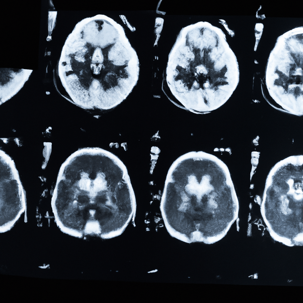
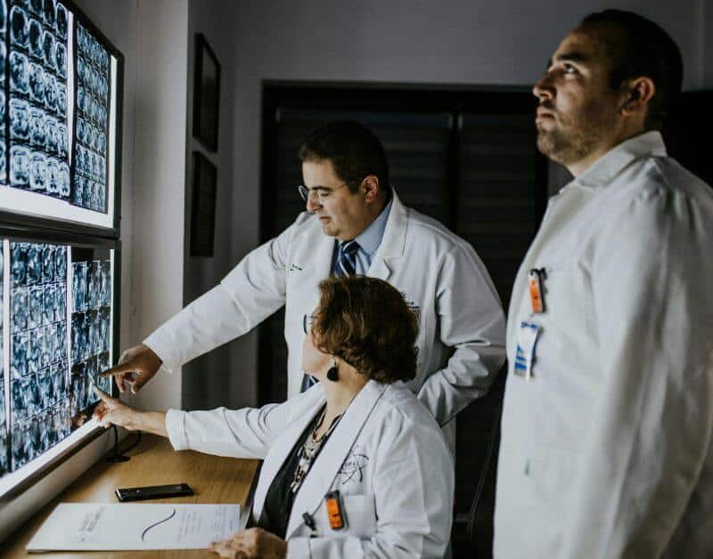
We have in-house Radiologist at MRI Chandigarh so, you will get same day report with accurate results.
We provide free pick and drop facility for our patients in tricity for MRI, CT Scan, PSMA PET Scan, DOTA PET Scan and PET Scan @ ₹12,999 in Chandigarh.
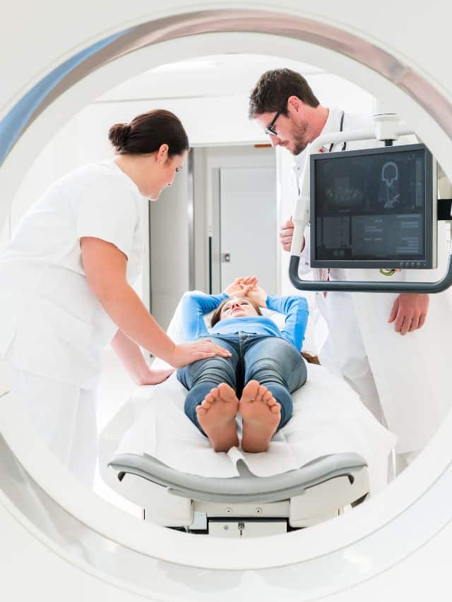
We are equipped with World Class MRI Scan Facility at Reasonable pricing. With best in class skilled technicians.
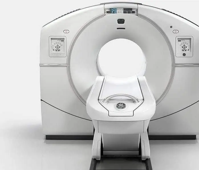
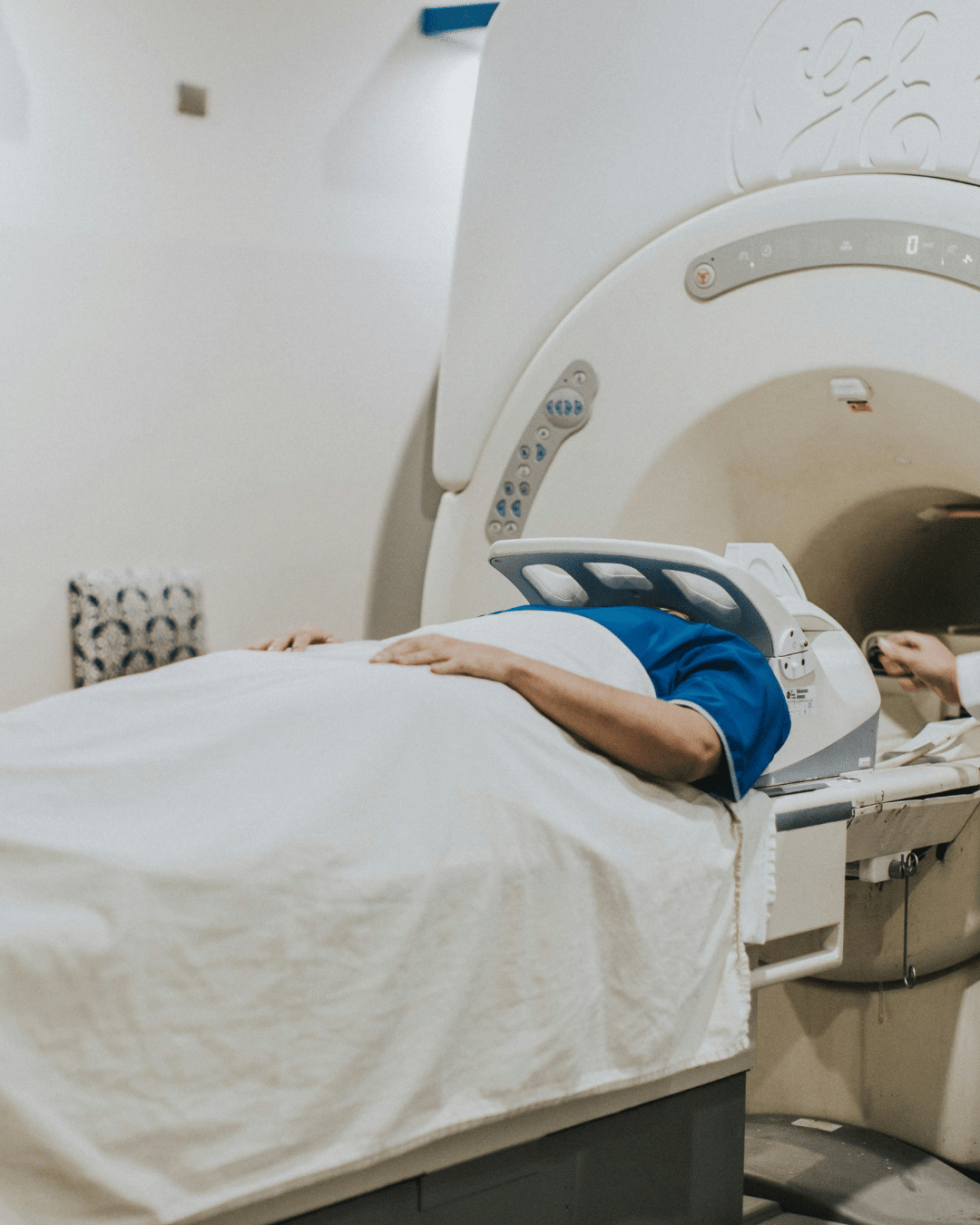
Get a comfortable MRI scan in Chandigarh using high-quality 1.5T and 3T machines with expert reporting and transparent, patient-friendly pricing.
MRI Chandigarh is your one-stop destination for advanced medical imaging and diagnostic services in Chandigarh. As one of the most trusted MRI centres in Chandigarh, we provide cutting-edge radio-imaging solutions, including both 1.5 Tesla and 3 Tesla MRI scans, at highly competitive prices—without compromising on quality.
We offer quick, accurate, and affordable MRI scans in Chandigarh with no hidden charges. Our experienced radiologists ensure timely results and, if needed, offer recommendations for further medical consultation based on your MRI report.
The cost of an MRI scan in Chandigarh varies depending on the type of scan and the diagnostic center. Prices typically start from ₹3,800 to ₹5,500. For example, 1.5 Tesla MRI scans may begin at ₹3,800, while 3 Tesla MRI scans are priced higher due to their advanced imaging capabilities. It's advisable to contact local MRI centers for precise pricing.
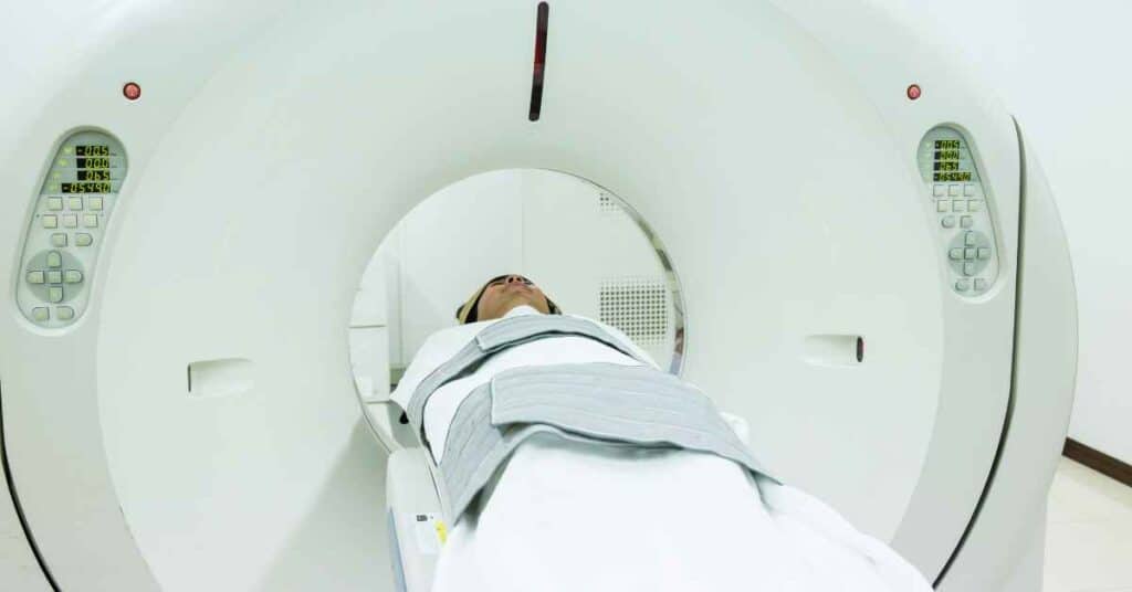
Preparation for an MRI scan generally involves minimal steps. For most MRI scans, you can maintain your regular diet. However, for abdominal scans, fasting for 3-4 hours prior to the procedure may be required. Wear loose-fitting, metal-free clothing, and inform your radiologist about any existing medical conditions, allergies, or implants.
Yes, MRI scans are generally considered safe as they do not use ionizing radiation. However, patients with certain implants like pacemakers should inform their doctor beforehand. It's crucial to remove all metal objects before the scan to prevent any risks associated with the MRI's strong magnetic field.
The duration of an MRI scan depends on the area being examined but typically ranges from 15 minutes to over an hour. For instance, a brain MRI may take about 30 to 60 minutes, while joint MRIs like knee or shoulder can take 15 to 45 minutes. Advanced 3 Tesla MRI machines can perform scans faster, enhancing patient comfort.
Chandigarh hosts several reputable MRI centres equipped with state-of-the-art technology and experienced radiologists. Centers like MRI Chandigarh are known for their advanced imaging services, including 1.5 Tesla and 3 Tesla MRI scans, and patient-centric care. It's recommended to research and choose a center that best fits your diagnostic needs.
While some diagnostic centers may allow self-referred MRI scans, it's advisable to consult with a healthcare professional to ensure the appropriateness of the scan and to provide the necessary clinical context for accurate interpretation.
Yes, certain MRI centres in Chandigarh offer complimentary pick-up and drop-off services within the Tricity area (Chandigarh, Mohali, Panchkula) to enhance patient convenience. It's best to inquire with the specific MRI center about the availability of such services. We provide free pick and drop in tricity for all scans including CT Scan & PET Scan too.
We are the leading diagnostic centre in Chandigarh providing comprehensive radio diagnostic services including MRI, CT Scan, PET Scan, Ultrasound, X-ray, Echocardiogram, ECG, and EEG at affordable rates with same-day reporting.
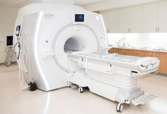
Experience the most comfortable and advanced MRI scans in Chandigarh with our high-field strength 1.5 Tesla and 3.0 Tesla machines. Our state-of-the-art MRI technology delivers exceptional quality diagnostic images for accurate results. Located in Sector 34, we provide the most reliable MRI services in Chandigarh Tricity at affordable rates.
Our Chandigarh diagnostic center provides world-class CT Scan facilities at the most competitive prices in the tricity region. Equipped with low-radiation dose CT scanners and operated by skilled technicians, we ensure the safest and most accurate CT scan services in Chandigarh, Mohali and Panchkula area.
Get the most affordable PET scan in Chandigarh at just Rs.16,000, the lowest price in the tricity region. Our all-inclusive package offers free pickup and drop, complimentary meals, and patient comfort amenities. Our Chandigarh PET scan center uses state-of-the-art technology for early cancer detection and treatment monitoring.
Our Chandigarh diagnostic center offers high-precision ultrasound services including 2D, 3D, and 4D scans. We use latest technology equipment at our partner diagnostic facilities across Chandigarh Sectors 34, 35, and 22. Our experienced radiologists provide accurate results for abdominal, pregnancy, thyroid, and other specialized ultrasound examinations.
Our network of partner X-ray centers in Chandigarh offers affordable digital X-ray services with instant results. Using advanced digital radiography technology, we provide high-quality bone and soft tissue imaging with minimal radiation exposure. Visit our X-ray diagnostic facilities in Sectors 34, 22, and 17 of Chandigarh for quick and reliable service.

Our diagnostic center in Chandigarh provides comprehensive cardiac and neurological testing including Echocardiography (Echo), Electrocardiogram (ECG), and Electroencephalogram (EEG). We offer the most affordable rates for these specialized tests in the Chandigarh region with reports prepared by experienced cardiologists and neurologists for accurate diagnosis.
As the leading diagnostic imaging center in Chandigarh, we provide comprehensive radiology services including MRI, CT Scan, PET-CT, Ultrasound, X-ray, Echocardiography, ECG, and EEG. Our state-of-the-art facilities are equipped with the latest diagnostic technology to deliver accurate results for patients across Chandigarh, Mohali, and Panchkula.
Visit our centers in Sector 34, Sector 22, or Sector 17 Chandigarh for the best diagnostic experience. Book your appointment today for high-quality, affordable diagnostic services in Chandigarh.
Positron Emission Tomography–Computed Tomography, commonly known as PET-CT, is an advanced hybrid imaging modality that plays a central role in modern diagnostic medicine.
It is widely used in oncology, cardiology, and neurology for disease detection, staging, treatment planning, and response assessment.
Continuous technological advancements have significantly enhanced the performance of PET-CT systems, resulting in improved image quality, higher diagnostic accuracy, and increased patient safety.
The latest PET-CT scan technology integrates digital detector systems, time-of-flight measurements, artificial intelligence–assisted image reconstruction, and optimized radiation dose management to support precise and reliable clinical evaluation.
PET-CT combines functional and anatomical imaging in a single examination. The PET component evaluates metabolic and molecular activity by detecting the distribution of radiotracers administered to the patient, most commonly fluorodeoxyglucose.
Tissues with increased metabolic demand demonstrate higher tracer uptake, allowing visualization of pathological processes at the cellular level.
The CT component provides high-resolution anatomical images that enable accurate localization of functional abnormalities.
The fusion of metabolic and structural information allows clinicians to correlate biological activity with precise anatomical detail, enhancing diagnostic confidence.
Conventional imaging modalities such as radiography, ultrasound, and standalone computed tomography primarily assess structural changes within tissues and organs.
While these techniques are essential in routine diagnostics, they may not detect early disease processes that manifest initially as metabolic or functional alterations.
Many pathological conditions, particularly malignancies, exhibit changes in cellular metabolism before structural abnormalities become apparent.
PET-CT addresses this limitation by identifying functional abnormalities at an early stage, allowing timely diagnosis and improved clinical management.
Early PET-CT systems relied on analog detector technology with limited sensitivity and temporal resolution. These systems were associated with longer acquisition times, higher image noise, and increased radiation exposure.
Recent advancements have led to the development of PET-CT scanners with digital detector architecture, enhanced signal processing, and advanced computational algorithms.
These improvements have resulted in superior spatial resolution, increased lesion detectability, reduced scan duration, and optimized radiation dose delivery, aligning PET-CT imaging with contemporary clinical standards.
The introduction of fully digital detector systems represents a major advancement in PET-CT technology. Digital detectors directly convert scintillation events into digital signals, providing improved timing resolution and higher sensitivity compared to analog systems. This allows accurate detection of low-level radiotracer uptake, facilitating identification of small or early-stage lesions. Enhanced detector efficiency also enables high-quality imaging with lower radiotracer doses, contributing to improved patient safety without compromising diagnostic performance.
Time-of-flight technology enhances PET image reconstruction by measuring the precise difference in arrival times of gamma photons generated during positron annihilation. Incorporation of this temporal information allows more accurate localization of the annihilation event along the line of response. The clinical benefits include improved contrast resolution, reduced image noise, and shorter acquisition times. These advantages are particularly relevant in patients with larger body habitus and in anatomically complex regions where image degradation was previously a challenge.
Artificial intelligence has become an integral component of contemporary PET-CT imaging workflows. Advanced reconstruction algorithms improve image quality by reducing noise, enhancing spatial resolution, and standardizing quantitative measurements. These techniques enable diagnostically reliable images to be obtained from lower-count data, supporting radiation dose reduction strategies. Artificial intelligence tools also assist in lesion detection, segmentation, and quantitative analysis, contributing to improved consistency and accuracy in image interpretation.
Radiation exposure remains an important consideration in diagnostic imaging. The latest PET-CT systems incorporate multiple strategies to minimize radiation dose while maintaining diagnostic accuracy. These include high-sensitivity digital detectors, adaptive dose modulation, and advanced reconstruction techniques. Modern PET-CT scanners achieve high-quality imaging with significantly lower radiation exposure compared to earlier generations. This is particularly important for pediatric patients, individuals undergoing repeated follow-up examinations, and patients requiring long-term disease surveillance.
Physiological motion, particularly respiratory movement, can adversely affect image quality in PET-CT studies of the thorax and upper abdomen. Contemporary PET-CT scanners incorporate motion correction and respiratory gating techniques that synchronize image acquisition with the patient’s breathing cycle. These approaches reduce motion-related artifacts, improve lesion delineation, and enhance quantitative accuracy. Improved motion management is essential for reliable evaluation of pulmonary, hepatic, and mediastinal lesions.
Modern PET-CT systems offer extended axial field-of-view capabilities, allowing larger anatomical regions to be imaged simultaneously. This increases system sensitivity and reduces overall scan time, enabling comprehensive whole-body imaging in a single session. Extended coverage enhances detection of metastatic disease and improves staging accuracy in oncology patients. Reduced acquisition time also improves patient comfort and operational efficiency in clinical practice.
Although fluorodeoxyglucose remains the most widely used PET radiotracer, advances in radiopharmaceutical development have expanded the clinical applications of PET-CT. Modern systems support a range of disease-specific radiotracers that target particular molecular pathways or receptor systems. These tracers are used in the evaluation of prostate cancer, neuroendocrine tumors, neurological disorders, and cardiovascular disease. Molecular imaging with PET-CT provides insights into disease biology that extend beyond anatomical assessment, supporting individualized diagnostic evaluation.
PET-CT is a fundamental tool in oncological imaging, providing critical information for diagnosis, staging, treatment planning, and response assessment. By identifying metabolically active tumor tissue, PET-CT enables differentiation between benign and malignant lesions with high accuracy. Accurate staging facilitates appropriate therapeutic selection and helps avoid unnecessary interventions. Advanced PET-CT technology improves detection of nodal involvement and distant metastases, contributing to comprehensive disease assessment.
PET-CT is particularly valuable for evaluating treatment response, as changes in metabolic activity often occur before structural changes become evident. Quantitative PET parameters allow objective assessment of therapeutic effectiveness and early identification of inadequate response. The reproducibility of measurements provided by modern PET-CT systems supports reliable longitudinal monitoring and early detection of disease recurrence, enabling timely clinical intervention.
Beyond oncology, PET-CT has established roles in cardiology and neurology. In cardiac imaging, PET-CT is used to assess myocardial perfusion, viability, and inflammatory conditions. In neurology, PET-CT contributes to the evaluation of epilepsy, cognitive disorders, and neurodegenerative diseases through analysis of cerebral metabolism and receptor activity. Advances in image resolution and quantitative accuracy have enhanced the clinical utility of PET-CT across these specialties.
Modern PET-CT scanners are designed to improve patient comfort and clinical workflow efficiency. Shorter scan times, automated acquisition protocols, and streamlined imaging processes reduce patient discomfort and improve compliance. Wider gantry openings and quieter system operation further enhance patient experience. Improved workflow efficiency supports timely image interpretation and clinical decision-making.
The diagnostic accuracy of PET-CT imaging depends on both the technological capabilities of the scanner and the expertise of the interpreting clinicians. Facilities equipped with advanced PET-CT systems and experienced nuclear medicine specialists are better positioned to provide accurate and clinically meaningful results. Adherence to standardized imaging protocols and quality assurance practices is essential for maintaining diagnostic reliability.
Ongoing research in PET-CT technology focuses on further expansion of field-of-view, development of novel radiotracers, and refinement of artificial intelligence applications. These advancements aim to enhance sensitivity, reduce radiation exposure, and provide comprehensive evaluation of systemic disease processes. Continued innovation is expected to further strengthen the role of PET-CT in precision diagnostic medicine.
The latest PET-CT scan technology represents a significant advancement in diagnostic imaging, combining digital detector precision, time-of-flight accuracy, artificial intelligence–assisted reconstruction, and optimized radiation dose management. These developments have enhanced the diagnostic capabilities of PET-CT across multiple clinical disciplines. By enabling early disease detection, accurate staging, reliable treatment monitoring, and comprehensive whole-body evaluation, modern PET-CT imaging supports informed clinical decision-making and improved patient care.


