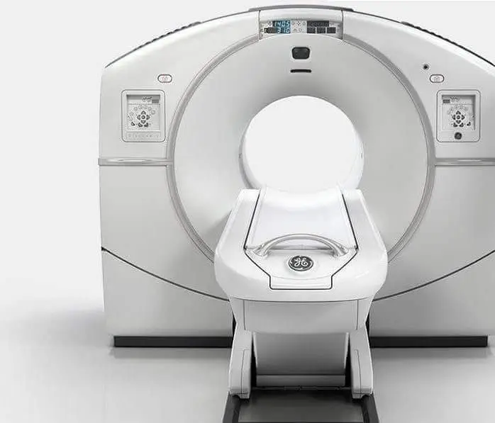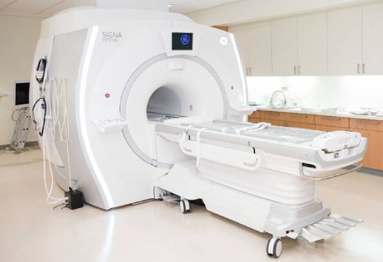


We have in-house Radiologist at MRI Chandigarh so, you will get same day report with accurate results.
We provide free pick and drop facility for our patients in tricity for MRI, CT Scan, PSMA PET Scan, DOTA PET Scan and PET Scan with cheapest price in Chandigarh.
We are equipped with World Class MRI Scan Facility at Reasonable pricing. With best in class skilled technicians.
Get the most comfortable MRI scan in Chandigarh with high quality 1.5T or 3T machines at best and affordable price.


MRI Chandigarh is your one-stop destination for advanced medical imaging and diagnostic services in Chandigarh. As one of the most trusted MRI centres in Chandigarh, we provide cutting-edge radio-imaging solutions, including both 1.5 Tesla and 3 Tesla MRI scans, at highly competitive prices—without compromising on quality.
We offer quick, accurate, and affordable MRI scans in Chandigarh with no hidden charges. Our experienced radiologists ensure timely results and, if needed, offer recommendations for further medical consultation based on your MRI report.
The cost of an MRI scan in Chandigarh varies depending on the type of scan and the diagnostic center. Prices typically start from ₹2,800 to ₹5,500. For example, 1.5 Tesla MRI scans may begin at ₹2,800, while 3 Tesla MRI scans are priced higher due to their advanced imaging capabilities. It's advisable to contact local MRI centers for precise pricing.
Preparation for an MRI scan generally involves minimal steps. For most MRI scans, you can maintain your regular diet. However, for abdominal scans, fasting for 3-4 hours prior to the procedure may be required. Wear loose-fitting, metal-free clothing, and inform your radiologist about any existing medical conditions, allergies, or implants.
Yes, MRI scans are generally considered safe as they do not use ionizing radiation. However, patients with certain implants like pacemakers should inform their doctor beforehand. It's crucial to remove all metal objects before the scan to prevent any risks associated with the MRI's strong magnetic field.
The duration of an MRI scan depends on the area being examined but typically ranges from 15 minutes to over an hour. For instance, a brain MRI may take about 30 to 60 minutes, while joint MRIs like knee or shoulder can take 15 to 45 minutes. Advanced 3 Tesla MRI machines can perform scans faster, enhancing patient comfort.
Chandigarh hosts several reputable MRI centres equipped with state-of-the-art technology and experienced radiologists. Centers like MRI Chandigarh are known for their advanced imaging services, including 3 Tesla MRI scans, and patient-centric care. It's recommended to research and choose a center that best fits your diagnostic needs.
While some diagnostic centers may allow self-referred MRI scans, it's advisable to consult with a healthcare professional to ensure the appropriateness of the scan and to provide the necessary clinical context for accurate interpretation.
Yes, certain MRI centres in Chandigarh offer complimentary pick-up and drop-off services within the Tricity area (Chandigarh, Mohali, Panchkula) to enhance patient convenience. It's best to inquire with the specific MRI center about the availability of such services. We provide free pick and drop in tricity for all scans including CT Scan & PET Scan too.
We are the leading diagnostic centre in Chandigarh providing comprehensive radio diagnostic services including MRI, CT Scan, PET Scan, Ultrasound, X-ray, Echocardiogram, ECG, and EEG at affordable rates with same-day reporting.

Experience the most comfortable and advanced MRI scans in Chandigarh with our high-field strength 1.5 Tesla and 3.0 Tesla machines. Our state-of-the-art MRI technology delivers exceptional quality diagnostic images for accurate results. Located in Sector 34, we provide the most reliable MRI services in Chandigarh Tricity at affordable rates.

Our Chandigarh diagnostic center provides world-class CT Scan facilities at the most competitive prices in the tricity region. Equipped with low-radiation dose CT scanners and operated by skilled technicians, we ensure the safest and most accurate CT scan services in Chandigarh, Mohali and Panchkula area.

Get the most affordable PET scan in Chandigarh at just Rs.16,000, the lowest price in the tricity region. Our all-inclusive package offers free pickup and drop, complimentary meals, and patient comfort amenities. Our Chandigarh PET scan center uses state-of-the-art technology for early cancer detection and treatment monitoring.

Our Chandigarh diagnostic center offers high-precision ultrasound services including 2D, 3D, and 4D scans. We use latest technology equipment at our partner diagnostic facilities across Chandigarh Sectors 34, 35, and 22. Our experienced radiologists provide accurate results for abdominal, pregnancy, thyroid, and other specialized ultrasound examinations.

Our network of partner X-ray centers in Chandigarh offers affordable digital X-ray services with instant results. Using advanced digital radiography technology, we provide high-quality bone and soft tissue imaging with minimal radiation exposure. Visit our X-ray diagnostic facilities in Sectors 34, 22, and 17 of Chandigarh for quick and reliable service.

Our diagnostic center in Chandigarh provides comprehensive cardiac and neurological testing including Echocardiography (Echo), Electrocardiogram (ECG), and Electroencephalogram (EEG). We offer the most affordable rates for these specialized tests in the Chandigarh region with reports prepared by experienced cardiologists and neurologists for accurate diagnosis.
As the leading diagnostic imaging center in Chandigarh, we provide comprehensive radiology services including MRI, CT Scan, PET-CT, Ultrasound, X-ray, Echocardiography, ECG, and EEG. Our state-of-the-art facilities are equipped with the latest diagnostic technology to deliver accurate results for patients across Chandigarh, Mohali, and Panchkula.
Visit our centers in Sector 34, Sector 22, or Sector 17 Chandigarh for the best diagnostic experience. Book your appointment today for high-quality, affordable diagnostic services in Chandigarh.
The knee MRI utilizes a strong electromagnetic field, radiofrequency, and computer to create detailed imagery of the components inside the knee joints. It is usually utilized in and around the joint to assist diagnose or assess discomfort, weakness, edema, or hemorrhage. Knee MRI doesn’t really utilize ionizing radiation and may assist in determining if surgery is needed.
Detailed pictures of the components inside the knee construction from different perspectives include bones, ligaments, nerves, joints, tendons plus blood vessels. MRI is just a test physician used for the diagnosis of medical problems. MRI is a non-intrusive test. Detailed pictures of MR let physicians to study bodily conditions and diagnose illness.
For 2 factors, the knee has become one of the body’s very often damaged sections. It works as a carrier of load, as well as the hip and ankles congruency does not demonstrate stability.
A comprehensive examination should be carried out in order to deal with problems like an acutely inflamed knee in a typical knee pathology appearance to ensure a correct diagnosis that can be followed by a correct treatment strategy. Magnetic resonance imaging, generally called as MRI, is generally regarded as one of the simplest yet non-invasive screening procedures.
In some cases, physicians will additionally suggest knee arthroscopy using an MRI, an operating technique used to diagnose and correct problems within knee joints. The etiology of the knee, as well as its components, is shown by the MR arthrogram throughout this procedure. Like a scientific study describes, MR arthrography is usually used to measure meniscus injuries inside the knee after the operation, and for monitoring lesions or malignancies in the knees bones.
Patients above the age of 65 usually experience neurodegenerative alterations or meniscal rupture patellofemoral (described as “runners knee”). Such signs can be diagnosed by clinical sensitivity testing. Furthermore, MRIs with simple radiograms might be suggested in certain circumstances in order to correctly establish the source of probable knee problems. MR images are capable of tracking rips, lumps, or recurring anterior knee discomfort in individuals less than 60 years of age.
Studies have already demonstrated that MRI performs an important part in the assessment of knee injuries. An MRI can be needed in order to further examine the lesion following a thorough first examination of the physical structure as well as an examination of the medical history of the individual. Some individuals may be subject to malignancies and tumor-like diseases due to the synovial membrane tissue lined with joints including the knee. The 2016 study termed MRI the ‘best and ideal method’ for providing greater details on the scope of such knee injuries. The method of imaging was also effective in identifying tumors from the other masses inside the joint region.
An MRI does not utilize radiation, in contrast to x-rays as well as CT scans. For everybody, particularly youngsters and women who are pregnant, this is generally a better option. CT scans rely on radiation amounts that are acceptable for grownups, but this is less safe for the children.
You confront various dangers when you have metal-containing implants. MRI magnets will create pacemaker issues.
Several individuals may experience an allergic response to the contrasting color during an MRI. Gadolinium is by far the most prevalent kind of contrast material. These allergic responses are generally minor and readily managed via medicine, based on the Radiological Society of North America.
The facility is clean, the staff is courteous, and the reports are provided in a timely fashion. All in all a great place for MRI scan in Chandigarh.
Staff is friendly I am happy centre is cleaned. Staff care the patient it is very good diagnostic centre
Used their service recently for my brother's MRI in Chandigarh and they had made it very convenient to book the scan and not just that they even told me what prerequisites I needed to take like fasting overnight. Will definitely use their service again.
Tried them for the first time for my MRI scan. Great service over the phone for appointment and from the technician who performed the scan. Got the report the same day by evening. Great experience overall.
I have got three scans done in the last few months at this centre. I recommend MRI Chandigarh to my family and friends for MRI scan if there is a need. For me most importantly the technician are very capable. Also overall not a bad experience at all. I would easily recommend this place to people i know.
3T MRI Scan in Chandigarh - 3T MRI centre is best because the fees of MRI is less as compare to all MRI centre in Chandigarh which is near by PGI. I done my MRI and the report is pretty accurate.
Fantastic experience. Everything is so managed here that you will not face any single problem. Staff members are also very polite. Recommended!
My 3rd PET CT has been done here. Really good services. Most of staff is very caring and polite. If you want to excellent results in same day, this PET CT is good option.
Had really great experience with Mri Scan in Chandigarh. The staff was really cooperative and helped me on my every visit. Doctors are really polite and guided me through. My scan went really smooth. Thank you for the best healthcare.