Best Diagnostic Center in Chandigarh for All Medical Imaging Needs
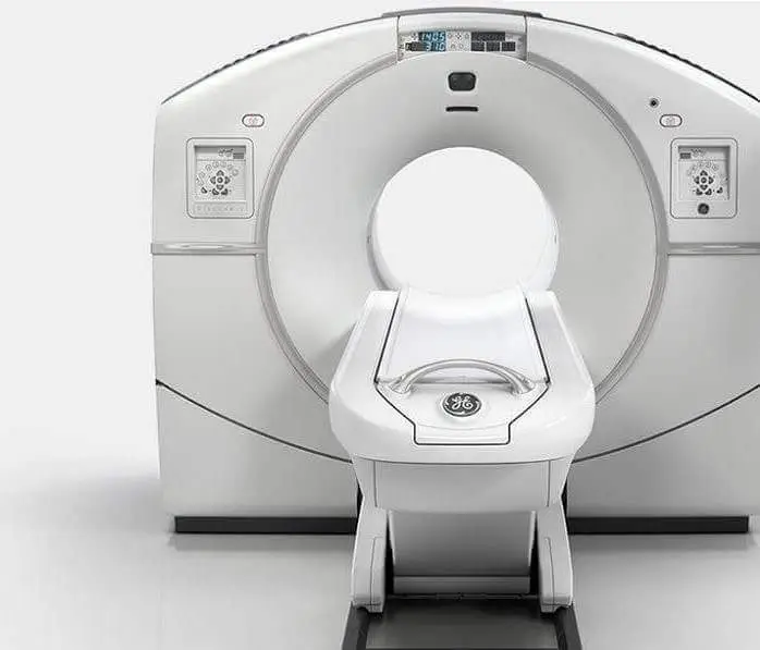
As the leading diagnostic imaging center in Chandigarh, we provide comprehensive radiology services including MRI, CT Scan, PET-CT, Ultrasound, X-ray, Echocardiography, ECG, and EEG. Our state-of-the-art facilities are equipped with the latest diagnostic technology to deliver accurate results for patients across Chandigarh, Mohali, and Panchkula.
Why Choose Our Diagnostic Services in Chandigarh?
- Advanced technology with 1.5T and 3.0T MRI scanners
- Most affordable rates for all diagnostic tests in Chandigarh Tricity
- Same-day reporting by specialized radiologists
- Conveniently located centers across Chandigarh
- Complimentary pick and drop services for patients
- Open 7 days a week including holidays
Visit our centers in Sector 34, Sector 22, or Sector 17 Chandigarh for the best diagnostic experience. Book your appointment today for high-quality, affordable diagnostic services in Chandigarh.
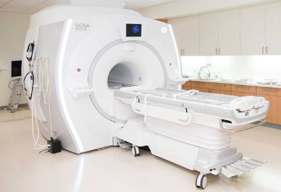
A Spinal MRI is used to produce a crisp and detailed image of the spinal cord with strong magnets, electromagnetic waves plus computers. This scan may be needed to monitor spinal issues, including lower back discomfort, neck discomfort, numbness, cramping, and stiffness in legs as well as arms.
The MRI will examine all or just portions of the spine. In contrast to X-rays and CT scans, harmful radiation does not occur. This procedure is secure and simple in general. You will be informed by the doctor of any potential hazards. You will also know whether the surgery is not possible due to some implants.
Why does someone need a spinal MRI?
After an accident in the spine, you will acquire an MRI. This is a scenario of urgency. The MRI also allows your physician to check the so-called tiny bones, the vertebras that compose your column, as well as the spinal discs, backbone, and spinal cord.
The assessment looks for: Spinal cord issues, tumors, odd elements in the backbone, bends in the spinal column, vertebrae breakage, wounds, inflammation, and swelling.
You may require a spinal MRI when you experience any of the following symptoms:
- Ache in the bottom back.
- Pain in the neck.
- Mid-back discomfort.
- Arms as well as chest radiate pain.
- Pain that radiates to the legs.
- Rigidity in your lower spine limits the range of movement.
- Due to stiffness, you cannot keep a regular posture.
- Muscular Cramps during exercise.
- Pain lasts from ten to fourteen days.
- Absence of motor foot function.
- Lack of urine or stool control.
Risks of Spinal MRI
MRI examinations are better than Computed tomography or X-ray examinations. But there are hazards, though. A magnet creating a powerful magnetic field is used by MRI. This can lead to difficulties like:
- Pulling medical equipment or moving it into the body.
- For MRI the device gets hot, which can burn any part.
- Causing malfunctioning of electrically powered medical equipment.
- Injury caused by the projectiles of magnetic objects.
The existence of medical equipment might potentially influence the MRI picture quality. The physician will verify in advance whether or not you have any equipment. After assessing your body if it is ok then you may get a scan.
Devices, not MRI compatible include:
Implants including pacemakers, auditory devices, prosthetic joints as well as stents, when you possess one of these, the equipment model may be asked by your physician or the MRI technician to check if an MRI will be appropriate for you.
Objects of metal, for example, operational clips, flatbeds, screws or cord mesh. When you possess these you might not be allowed to have an MRI on that region of your body.
Other risks of MRI include:
Claustrophobia- Some people experience this because they have to enter the MRI tunnel or tube. To aid you or attempt an open MRI scanner, you will consume anti-anxiety medicine in advance.
Allergic reactivity or dye-contrast sensitivity- Physicians may utilise MRI dyes. Certain individuals are allergic to dyes and should not undergo MRIs.
Conclusion
A professionally educated physician named a radiologist will examine your MRI of the spine and tell your doctor about the findings. What they signify and what they should do next is to be explained by your physician. To obtain your findings, it may require up to one week or longer.


