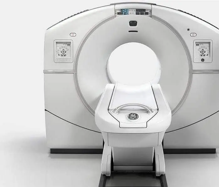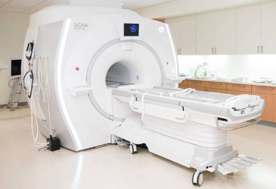


We have in-house Radiologist at MRI Chandigarh so, you will get same day report with accurate results.
We provide free pick and drop facility for our patients in tricity for MRI, CT Scan, PSMA PET Scan, DOTA PET Scan and PET Scan with cheapest price in Chandigarh.
We are equipped with World Class MRI Scan Facility at Reasonable pricing. With best in class skilled technicians.
Get the most comfortable MRI scan in Chandigarh with high quality 1.5T or 3T machines at best and affordable price.


MRI Chandigarh is your one-stop destination for advanced medical imaging and diagnostic services in Chandigarh. As one of the most trusted MRI centres in Chandigarh, we provide cutting-edge radio-imaging solutions, including both 1.5 Tesla and 3 Tesla MRI scans, at highly competitive prices—without compromising on quality.
We offer quick, accurate, and affordable MRI scans in Chandigarh with no hidden charges. Our experienced radiologists ensure timely results and, if needed, offer recommendations for further medical consultation based on your MRI report.
The cost of an MRI scan in Chandigarh varies depending on the type of scan and the diagnostic center. Prices typically start from ₹2,800 to ₹5,500. For example, 1.5 Tesla MRI scans may begin at ₹2,800, while 3 Tesla MRI scans are priced higher due to their advanced imaging capabilities. It's advisable to contact local MRI centers for precise pricing.
Preparation for an MRI scan generally involves minimal steps. For most MRI scans, you can maintain your regular diet. However, for abdominal scans, fasting for 3-4 hours prior to the procedure may be required. Wear loose-fitting, metal-free clothing, and inform your radiologist about any existing medical conditions, allergies, or implants.
Yes, MRI scans are generally considered safe as they do not use ionizing radiation. However, patients with certain implants like pacemakers should inform their doctor beforehand. It's crucial to remove all metal objects before the scan to prevent any risks associated with the MRI's strong magnetic field.
The duration of an MRI scan depends on the area being examined but typically ranges from 15 minutes to over an hour. For instance, a brain MRI may take about 30 to 60 minutes, while joint MRIs like knee or shoulder can take 15 to 45 minutes. Advanced 3 Tesla MRI machines can perform scans faster, enhancing patient comfort.
Chandigarh hosts several reputable MRI centres equipped with state-of-the-art technology and experienced radiologists. Centers like MRI Chandigarh are known for their advanced imaging services, including 3 Tesla MRI scans, and patient-centric care. It's recommended to research and choose a center that best fits your diagnostic needs.
While some diagnostic centers may allow self-referred MRI scans, it's advisable to consult with a healthcare professional to ensure the appropriateness of the scan and to provide the necessary clinical context for accurate interpretation.
Yes, certain MRI centres in Chandigarh offer complimentary pick-up and drop-off services within the Tricity area (Chandigarh, Mohali, Panchkula) to enhance patient convenience. It's best to inquire with the specific MRI center about the availability of such services. We provide free pick and drop in tricity for all scans including CT Scan & PET Scan too.
We are the leading diagnostic centre in Chandigarh providing comprehensive radio diagnostic services including MRI, CT Scan, PET Scan, Ultrasound, X-ray, Echocardiogram, ECG, and EEG at affordable rates with same-day reporting.

Experience the most comfortable and advanced MRI scans in Chandigarh with our high-field strength 1.5 Tesla and 3.0 Tesla machines. Our state-of-the-art MRI technology delivers exceptional quality diagnostic images for accurate results. Located in Sector 34, we provide the most reliable MRI services in Chandigarh Tricity at affordable rates.

Our Chandigarh diagnostic center provides world-class CT Scan facilities at the most competitive prices in the tricity region. Equipped with low-radiation dose CT scanners and operated by skilled technicians, we ensure the safest and most accurate CT scan services in Chandigarh, Mohali and Panchkula area.

Get the most affordable PET scan in Chandigarh at just Rs.16,000, the lowest price in the tricity region. Our all-inclusive package offers free pickup and drop, complimentary meals, and patient comfort amenities. Our Chandigarh PET scan center uses state-of-the-art technology for early cancer detection and treatment monitoring.

Our Chandigarh diagnostic center offers high-precision ultrasound services including 2D, 3D, and 4D scans. We use latest technology equipment at our partner diagnostic facilities across Chandigarh Sectors 34, 35, and 22. Our experienced radiologists provide accurate results for abdominal, pregnancy, thyroid, and other specialized ultrasound examinations.

Our network of partner X-ray centers in Chandigarh offers affordable digital X-ray services with instant results. Using advanced digital radiography technology, we provide high-quality bone and soft tissue imaging with minimal radiation exposure. Visit our X-ray diagnostic facilities in Sectors 34, 22, and 17 of Chandigarh for quick and reliable service.

Our diagnostic center in Chandigarh provides comprehensive cardiac and neurological testing including Echocardiography (Echo), Electrocardiogram (ECG), and Electroencephalogram (EEG). We offer the most affordable rates for these specialized tests in the Chandigarh region with reports prepared by experienced cardiologists and neurologists for accurate diagnosis.
As the leading diagnostic imaging center in Chandigarh, we provide comprehensive radiology services including MRI, CT Scan, PET-CT, Ultrasound, X-ray, Echocardiography, ECG, and EEG. Our state-of-the-art facilities are equipped with the latest diagnostic technology to deliver accurate results for patients across Chandigarh, Mohali, and Panchkula.
Visit our centers in Sector 34, Sector 22, or Sector 17 Chandigarh for the best diagnostic experience. Book your appointment today for high-quality, affordable diagnostic services in Chandigarh.
Due to the intricate three-dimensional structure, the foot and knee are some of the toughest places to picture. Magnetic resonance imaging (MRI) has proven a useful technique in assessing feet and ankles issues in individuals with its 3d abilities. MRI is not bones scintigraphy but extremely accurate than ultrasonography and computer scanning. The articulating area and joint region are confined to the arthroscopy on the ankle. Using a comprehensive inspection that surpasses the capacities of all other existing procedures, an MRI provides a worldwide assessment of bones, muscles, joints, and other tissues.
In addition to a comprehensive history and medical examination, imaging can help diagnose a person with a feet or ankles injury and create a treatment plan. Ankle and foot scanning can be helpful in preventing or eliminating a disease following a feet or ankles trauma or if a condition with a moderate treatment has not advanced. There are three common perspectives for the radiograph for the bone disorder.
The AP image is shown via the talus as well as the distal tibia portion. The lateral image shows the joint of the distal tibia and talus and a side image of the tarsal bones and calcaneus. So, without overlays from the Fibula, the mortise image shows the talus as well as the distal tibia.
A stress imaging can be obtained to assess ligamentous laxity when fragility is detected. If sensitive tissue or swelling of the tissue is detected, an MRI or ultrasound diagnosis may be needed.
Hallux Valgus: Hallux Valgus is a malfunction in which the main toe has diverged laterally, and in the first metatarsal, a valgus angle is created for the 1st MTP joint. Since this is a knotty malfunction, the MRI is the preferred choice for Hallux Valgus diagnosis.
Lateral Ankle Sprain: 95 % of knee sprains have always been involved in the lateral ankle tendons. Images of a lateral sprain can be shown by injury cause, pain region, and appropriate special testing. The research examined the capacity to detect where the tears were and discovered that MRIs could properly identify the injuries in 93% of cases.
Knee sprains are quite frequent wounds. 20 to 40% of acute instability individuals suffer from related conditions, including talus chondral lesions, peroneal tears, or loose body conditions. These lesions can lead to morbidity following a straining of the knee. Diagnoses and treatments during reconstruction ligament surgery might seem ideal if conclusive data were not available.
In order for ankle stiffness abnormalities to be properly treated, they need to be adequately identified. An initial crucial stage is a comprehensive medical examination, although some abnormalities may be challenging to evaluate. Preoperative MRIs are one approach to diagnose concurrent problems. If your wound fails to cure and the region of the damage continually bleeds, your doctor may order an MRI.
Meanwhile, MRI scanning is also more accurate and expensive for the techniques. The Foot or Ankle MRI scan costs are according to market location and competition. The inclusion of additional picture centers often reduces the scan costs. In general, costs are around 4950-5000 at a unit of hospital imaging. Patients with cash payment should pay for the MRI scan at any point between 2500 and 5500.
The facility is clean, the staff is courteous, and the reports are provided in a timely fashion. All in all a great place for MRI scan in Chandigarh.
Staff is friendly I am happy centre is cleaned. Staff care the patient it is very good diagnostic centre
Used their service recently for my brother's MRI in Chandigarh and they had made it very convenient to book the scan and not just that they even told me what prerequisites I needed to take like fasting overnight. Will definitely use their service again.
Tried them for the first time for my MRI scan. Great service over the phone for appointment and from the technician who performed the scan. Got the report the same day by evening. Great experience overall.
I have got three scans done in the last few months at this centre. I recommend MRI Chandigarh to my family and friends for MRI scan if there is a need. For me most importantly the technician are very capable. Also overall not a bad experience at all. I would easily recommend this place to people i know.
3T MRI Scan in Chandigarh - 3T MRI centre is best because the fees of MRI is less as compare to all MRI centre in Chandigarh which is near by PGI. I done my MRI and the report is pretty accurate.
Fantastic experience. Everything is so managed here that you will not face any single problem. Staff members are also very polite. Recommended!
My 3rd PET CT has been done here. Really good services. Most of staff is very caring and polite. If you want to excellent results in same day, this PET CT is good option.
Had really great experience with Mri Scan in Chandigarh. The staff was really cooperative and helped me on my every visit. Doctors are really polite and guided me through. My scan went really smooth. Thank you for the best healthcare.