
Description
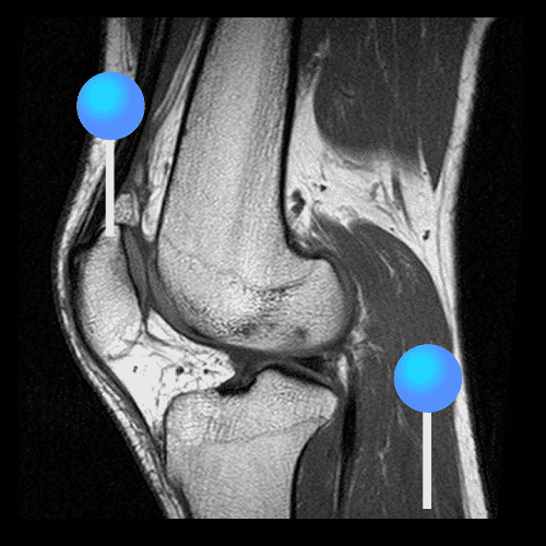
A Contrast CT Scan of the Knee is an advanced imaging test used to evaluate the knee joint, bones, cartilage, ligaments, muscles, and blood vessels. Because a contrast dye is used, this scan highlights soft tissues and abnormal areas, and therefore allows doctors to detect injuries, infections, and tumors with greater clarity.
Moreover, by combining high-resolution CT imaging with contrast enhancement, this test provides a more complete and accurate view of knee problems.
What Is a Contrast CT Knee Scan?
A contrast-enhanced CT of the knee uses X-ray technology and computer processing to produce detailed cross-sectional images. As a result, doctors can examine both bone and soft-tissue structures, which consequently improves diagnostic accuracy.
Why Is a Contrast CT of the Knee Performed?
This scan is recommended when symptoms are severe or unclear, and therefore it is used to:
- Therefore, detect ligament or cartilage injuries
- Additionally, identify tumors or abnormal growths
- Moreover, evaluate joint infections or inflammation
- Furthermore, assess blood vessel problems around the knee
- Finally, plan surgery or guide treatment
How Is the Scan Performed?
Before the scan, a contrast dye is injected into a vein. Meanwhile, you will lie on the CT table while images of the knee are taken. Consequently, the scanner produces clear, high-resolution pictures within minutes.
Most importantly, the test is painless and usually takes only 10 to 15 minutes.
Benefits of Contrast CT Knee Imaging
- Firstly, it provides detailed images of bones and soft tissues
- Secondly, it detects hidden injuries and disease
- Thirdly, it improves diagnostic confidence
- In addition, it supports accurate treatment planning
- Overall, it leads to better patient outcomes
Book a Contrast CT Knee Scan
If you have persistent knee pain, swelling, or injury, a Contrast CT Knee Scan not only clarifies the cause but also helps your doctor choose the most effective treatment.


Comments