
Description
A CT Scan of the Shoulder is an advanced diagnostic imaging test that uses X-rays and computer processing to create highly detailed cross-sectional images of the shoulder joint. It provides a much more precise and comprehensive view than standard X-rays, allowing doctors to evaluate both bone and soft-tissue structures.
This scan helps in identifying problems affecting the bones, joints, muscles, tendons, and surrounding tissues of the shoulder.
What Is a CT Shoulder Scan?
A CT shoulder scan captures multiple thin-slice images of the shoulder from different angles and combines them into detailed 3-dimensional views. This allows doctors to closely examine the internal structures of the shoulder.
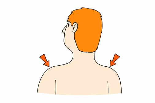
Why Is a CT Scan of the Shoulder Performed?
This test is commonly recommended to:
- Detect fractures or dislocations
- Evaluate shoulder pain or limited movement
- Assess joint damage or arthritis
- Identify tumors or infections
- Plan surgery or monitor healing
How Is the Scan Performed?
You will lie on a CT scanner table while the machine rotates around your shoulder to capture images. In some cases, contrast dye may be used to improve visibility of certain tissues.
The scan is painless and usually takes 10 to 15 minutes.
Benefits of CT Shoulder Imaging
- Provides detailed images of bone and joint structures
- Helps detect subtle injuries
- Fast and non-invasive
- Supports accurate diagnosis and treatment planning
Book a CT Scan of the Shoulder
If you have ongoing shoulder pain, injury, or abnormal X-ray findings, a CT scan of the shoulder offers a reliable way to identify the cause and guide proper treatment.


Comments