

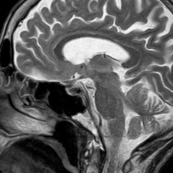
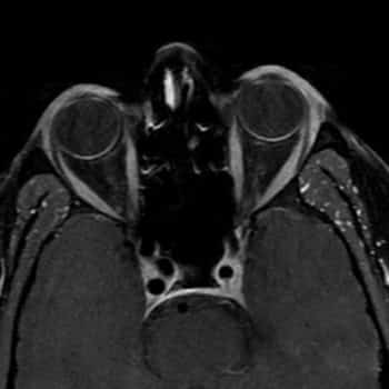
Description
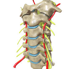
An MRI of the Whole Spine is a highly detailed imaging test that examines the cervical, thoracic, and lumbar regions of the spine in one complete scan. Because it uses magnetic fields instead of radiation, it provides clear and safe visualization of the spinal cord, nerves, discs, and surrounding tissues.
Moreover, by capturing precise cross-sectional images, this scan helps doctors identify spinal problems that may otherwise remain hidden.
What Is an MRI Whole Spine?
An MRI Whole Spine scan uses advanced imaging technology to create detailed pictures of the entire spinal column. As a result, it allows specialists to study the spine in its entirety rather than in isolated sections, which therefore improves diagnostic accuracy.
Why Is an MRI of the Whole Spine Performed?
This scan is recommended when symptoms involve more than one area of the spine or when the cause of pain is unclear. Consequently, it is used to:
- Therefore, detect disc bulges or herniation
- Additionally, identify nerve compression
- Moreover, evaluate spinal tumors or infections
- Furthermore, assess spinal cord disorders
- Finally, monitor degenerative spine disease
How Is the Scan Performed?
During the scan, you will lie comfortably on the MRI table. Meanwhile, the scanner captures detailed images of your entire spine. Consequently, doctors receive a complete picture of spinal health within a single session.
Most importantly, the test is painless and usually takes 45 to 60 minutes.
Benefits of MRI Whole Spine
- Firstly, it offers complete spinal assessment
- Secondly, it provides excellent soft-tissue detail
- Thirdly, it helps pinpoint the exact cause of pain
- In addition, it supports accurate treatment planning
- Overall, it improves long-term spine care
Book an MRI Whole Spine
If you are experiencing ongoing back pain, numbness, or weakness, an MRI Whole Spine not only identifies the problem but also helps your doctor choose the most effective treatment.


Comments