
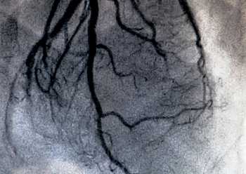
Description
A PET Scan for Cardiac Viability is a specialized heart imaging test used to determine whether areas of the heart muscle that are not functioning properly are still alive and capable of recovery. This advanced scan helps doctors decide whether treatments such as bypass surgery, angioplasty, or medication can improve heart function.
By measuring blood flow and glucose metabolism in the heart, cardiac viability PET imaging distinguishes between permanently damaged tissue and viable but weakened heart muscle.
What Is a Cardiac Viability PET Scan?
A cardiac viability PET scan uses a radioactive glucose tracer to evaluate how much energy different areas of the heart muscle are using. Healthy or recoverable heart tissue absorbs more tracer, while scarred or dead tissue shows little or no uptake.
Why Is a Cardiac Viability PET Scan Performed?
This scan is recommended for patients who have:
- Reduced heart pumping function
- History of heart attack
- Heart failure
- Blocked coronary arteries
- Consideration for bypass surgery or angioplasty
How Is the Test Performed?
A small amount of radiotracer is injected into a vein. The tracer travels to the heart muscle, and a PET scanner captures images showing glucose uptake and blood flow.
The scan is painless and typically takes 45 to 60 minutes.
Benefits of Cardiac Viability PET
- Identifies heart muscle that can recover with treatment
- Prevents unnecessary surgeries
- Helps guide the best treatment plan
- Provides highly accurate heart function assessment
Book a Cardiac Viability PET Scan
If you have heart disease or reduced heart function, a cardiac viability PET scan offers critical information to help your doctor choose the most effective treatment for improving heart health.

Comments