Hearing your doctor say that you need a PET/CT scan can immediately trigger a wave of anxiety. For many patients, the fear of the unknown-combined with intimidating medical terminology and the mention of “radioactive tracers”-makes the preparation leading up to the scan incredibly stressful.
However, a PET/CT scan is one of the most brilliant, life-saving advancements in modern medicine. Instead of just taking a static picture of your insides, this machine actually watches your cells function in real time.
If you or a loved one are preparing for this procedure, understanding exactly how the machine works takes the mystery out of the process. Here is the deep-dive science into what a PET/CT scan is, why it is used in oncology, and exactly how it detects cancer hiding in the human body.
Two Scans in One: Anatomy vs. Activity
To understand a PET/CT scan, you have to break down the name. It is actually two completely different scans performed simultaneously by one machine.
The CT (Computed Tomography) portion of the scan is basically a highly advanced, 3-dimensional X-ray. It takes precise pictures of your internal anatomy. It shows the doctor the exact size, shape, and location of your organs, bones, and any physical tumors. However, a CT scan cannot easily tell the difference between a cancerous tumor and a benign (harmless) cyst just by looking at the shape.
That is where the PET (Positron Emission Tomography) scan comes in. While the CT scan looks at your anatomy, the PET scan looks at your cellular metabolism (how actively your cells are eating and using energy). When combined, the doctor gets a complete map of both what your body looks like and how it is functioning.
The “Sugar” Secret: How Cancer Glows
The real magic of a PET scan doesn’t actually come from the machine itself; it comes from what happens about an hour before your scan begins.
Cancer cells share one major trait: they are incredibly greedy. Because cancer cells multiply at an abnormally fast rate, they require massive amounts of energy to survive. Their favorite source of energy is glucose (sugar).
To exploit this, medical professionals use a special liquid called FDG (Fluorodeoxyglucose). FDG is essentially a glucose (sugar) molecule attached to a safe, tiny amount of a radioactive isotope.
Before your scan, a nurse will inject this FDG tracer into your IV. You will then sit quietly in a dark room for about 60 minutes. During this time, the “radioactive sugar” travels through your bloodstream. Because cancer cells consume sugar much faster than healthy, normal cells, they absorb the majority of the FDG tracer.
When you finally lie down inside the PET/CT scanner, the machine detects the radiation emitted by the tracer. The cancer cells-stuffed full of the radioactive sugar-will literally “glow” or “light up” as bright spots on the doctor’s computer screen, exposing their exact location.
Why Oncologists Rely on PET/CT Scans?
Because the PET scan looks at cellular activity rather than just physical size, it is a game-changer for oncology in three major ways:
1. Early Detection: A PET scan can often detect biochemical changes in cells before a tumor is physically large enough to be seen on a standard MRI or CT scan.
2. Accurate Staging: By doing a full-body PET/CT scan, doctors can see if cancer cells have broken away from the original tumor and spread (metastasized) to other organs or lymph nodes.
3. Real-Time Treatment Monitoring: If you are undergoing chemotherapy or radiation, a PET scan can show if the treatment is working.
Even if a tumor hasn’t physically shrunk yet, the PET scan can reveal that the tumor is no longer glowing-meaning the cancer cells are dead and no longer consuming energy.
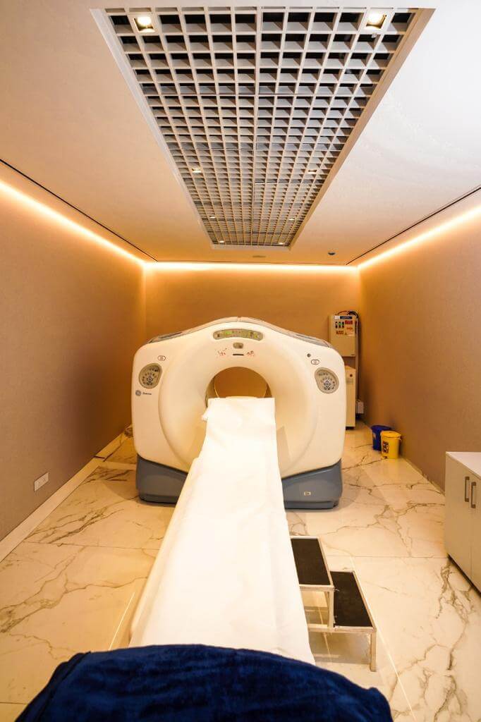
What to Expect on Scan Day?
Despite the high-tech science, the patient experience is remarkably easy and completely painless.
You will be asked to fast (eat nothing and drink only plain water) for about 6 hours before your appointment. This ensures your body’s natural blood sugar is very low, making the cancer cells “hungry” for the FDG tracer.
Once the tracer is injected and you have waited the required hour, you will lie flat on a motorized bed that slowly slides into a large, donut-shaped scanner. The scan itself usually takes between 20 to 40 minutes. You won’t feel anything, and the machine is much quieter than a traditional MRI. Afterward, you can drive yourself home, eat normally, and simply drink plenty of water to flush the remaining tracer out of your system.
A PET/CT scan is a remarkable window into the microscopic world of your biology. B
y understanding that the machine is simply using cancer’s own appetite against it to make it visible, you can walk into your appointment feeling empowered rather than afraid.
It is not just a test; it is the ultimate roadmap that helps your medical team design the exact plan needed to heal your body.

Subscribe
0 Comments

Comments