
Description
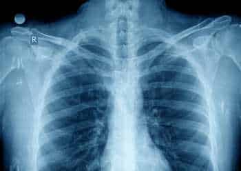
A Contrast CT Scan of the Shoulder is an advanced imaging test used to obtain detailed cross-sectional views of the shoulder joint, muscles, ligaments, tendons, and surrounding soft tissues. An iodine-based contrast dye is injected into a vein before the scan to enhance visibility of blood vessels and abnormal areas.
This scan helps doctors clearly identify injuries, inflammation, tumors, infections, and other conditions that may not be visible on routine X-rays.
What Is a Contrast CT Shoulder Scan?
A contrast-enhanced CT scan uses X-ray technology and computer processing to create high-resolution images of the shoulder. The contrast dye improves the clarity of soft tissues and blood vessels, allowing more accurate diagnosis.
Why Is a Contrast CT of the Shoulder Performed?
This scan is recommended to:
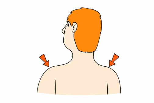
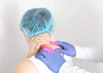
- Evaluate shoulder pain or limited movement
- Detect fractures, dislocations, or joint damage
- Identify tumors, infections, or inflammation
- Assess blood vessels around the shoulder
- Plan surgery or guide treatment
How Is the Scan Performed?
A contrast dye is injected into a vein, usually in the arm. You will lie on the CT scanner table while images of the shoulder are taken as the scanner rotates around you.
The procedure is painless and typically completed within 10 to 15 minutes.
Benefits of Contrast CT Shoulder Imaging
- Provides highly detailed images of bones and soft tissues
- Helps detect subtle abnormalities
- Quick and non-invasive
- Supports accurate diagnosis and treatment planning
Book a Contrast CT Shoulder Scan
If you have persistent shoulder pain, injury, or abnormal imaging findings, a Contrast CT Scan of the Shoulder offers precise and reliable information to help guide proper care.


Comments